

Dysfunctional uterine bleeding: symptoms and treatment
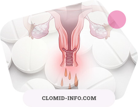
Dysfunctional uterine bleeding (DUB) is a form of menstrual dysfunction caused by a violation of the cyclic production of ovarian hormones. DUB can manifest as menorrhagia or menometrorrhagia. Functional changes leading to uterine bleeding can be at any level of regulation of menstrual function: in the cerebral cortex, hypothalamus, pituitary, adrenal glands, thyroid gland, ovaries. DUB recur and often lead to impaired reproductive function, and hormonal disorders in DUB - to the development of hyperplastic processes up to precancer and endometrial cancer.
Depending on the period of life, women emit:
- DUB juvenile period - 12-17 years;
- DUB of the reproductive period - 18-45 years;
- DUB of the premenopausal period - 46-55 years.
Reproductive dysfunctional uterine bleeding
DUB make up about 4-5% of gynecological diseases of the reproductive period and remain the most common pathology of the woman's reproductive system.
Etiology and pathogenesis. Etiological factors can be stressful situations, climate change, mental and physical fatigue, occupational hazards, adverse living conditions, hypovitaminosis, intoxication and infection, hormonal homeostasis disorders, abortion, and the use of certain medications. Along with the great importance of primary disturbances in the cortex-hypothalamus-pituitary system, primary disturbances at the level of the ovaries play an equally important role. The cause of ovulation disorders can be inflammatory and infectious diseases, under the influence of which a thickening of the ovarian protein membrane, a change in blood supply and a decrease in the sensitivity of ovarian tissue to gonadotropic hormones are possible.
Depending on the pathogenetic mechanisms and clinical and morphological features of the DUB of the reproductive period, they are divided into anovulatory and ovulatory.
In the reproductive period, the end result of hypothalamic-pituitary disorders is anovulation, which can be based on either persistence or atresia of the follicle. With DUB in the reproductive age in the ovaries, the follicle persists more often with an excess of estrogen production. Since ovulation does not occur and the corpus luteum does not form, a progesterone deficiency state is created and absolute hyperestrogenism occurs. The persistence of the follicle represents a kind of stoppage of the normal menstrual cycle in a period close to ovulation: the follicle, upon reaching maturity, does not undergo further physiological transformations, continuing to secrete estrogens. Anovulatory bleeding may be due to follicular atresia as a result of relative hyperestrogenia. In the ovary, one or more follicles stop at any stage of development, without undergoing further cyclic transformations, but without stopping functioning until a certain time. Subsequently, the atresizing follicles undergo reverse development or turn into small cysts. With atresia of follicles, there is little estrogen, but due to anovulation, the corpus luteum and the release of progesterone are absent - a state of relative hyperestrogenia develops.
Prolonged exposure to an elevated estrogen level on the uterus causes excessive growth of the endometrium. An increase in the duration and intensity of proliferative processes in the endometrium leads to hyperplasia with a risk of developing atypical hyperplasia and endometrial adenocarcinoma. Due to the lack of ovulation and the corpus luteum, there is not enough progesterone necessary for secretory transformation and normal rejection of the proliferative endometrium. The bleeding mechanism is associated with vascular changes: congestive plethora with a sharp expansion of capillaries in the endometrium, circulatory disorders, tissue hypoxia are accompanied by dystrophic changes in the uterine mucosa and the appearance of necrotic processes against the background of blood stasis and thrombosis. All of the above leads to a prolonged and uneven rejection of the endometrium. The morphological structure of the mucous membrane is mottled: along with areas of decay and rejection, there are foci of regeneration.
Ovulatory DUB is usually caused by persistence of the corpus luteum, which is more commonly observed at the age of over 30. The dysfunction of the corpus luteum lies in its long-term functional activity. As a result of persistence of the corpus luteum, the level of gestagens does not fall fast enough or remains at the same level for a long time. Uneven rejection of the functional layer leads to prolonged menometerorrhagia. A decrease in uterine tone under the influence of high progesterone in the blood also contributes to bleeding. In this case, the corpus luteum has no signs of reverse development, or along with luteal cells in a state of reverse development, there are areas with pronounced signs of functional activity. The persistence of the corpus luteum is evidenced by a high level of progesterone in the blood and an echographic picture of the ovaries.
During bleeding in the endometrium, the content of prostaglandin F2 is decreased, which enhances vascular contraction, and the content of prostaglandin E2 is increased, which prevents platelet aggregation.
Ovulatory bleeding can occur in the middle of the menstrual cycle, after ovulation. Normally, in the middle of the menstrual cycle, there is a slight decrease in estrogen levels, but it does not lead to bleeding, since the general hormonal level is maintained by the corpus luteum starting to function. With a significant and sharp decline in hormone levels after an ovulatory peak, blood discharge from the genital tract is observed for 2-3 days.
The clinical manifestations of dysfunctional uterine bleeding are usually determined by changes in the ovaries. The main complaint in patients with DUB is a violation of the rhythm of menstruation: bleeding often occurs after a delay in menstruation or menometorrhagia is noted. If the persistence of the follicle is short-term, then uterine bleeding in intensity and duration does not differ from normal menstruation. More often, the delay is quite long (up to 6-8 weeks), after which bleeding occurs. Often it begins as moderate, periodically decreases, intensifies again and lasts a very long time, leading to anemia and weakening of the body.
DUB due to persistence of the corpus luteum - menstruation that occurs on time or after a short delay. With each new cycle, it becomes longer and more plentiful, turning into a menometerorrhagia, lasting up to 1-1.5 months.
Impaired ovarian function in patients with DUB can lead to decreased fertility.
When diagnosing, it is necessary to exclude other causes of bleeding, which in reproductive age can be: benign and malignant diseases of the genital organs, endometriosis, uterine fibroids, genital injuries, inflammatory processes of the uterus and appendages, interrupted uterine and ectopic pregnancy, remnants of the fetal egg after an abortion or spontaneous miscarriage, placental polyp after childbirth or abortion. Uterine bleeding occurs with extragenital diseases: diseases of the blood, liver, cardiovascular system, endocrine pathology. The examination should be aimed at eliminating morphological pathology and determining functional disorders in the hypothalamus-pituitary-ovary-uterus system using generally available, and if necessary, additional examination methods. At the 1st stage after clinical methods (history, objective general and gynecological examination), hysteroscopy is performed with separate diagnostic curettage and morphological examination of scrapings. Subsequently, after stopping the bleeding, it is shown:
- laboratory research (clinical blood test, coagulogram) to assess anemia and the condition of the blood coagulation system;
- examination according to tests of functional diagnostics (measuring basal temperature, a symptom of a “pupil”, a symptom of tension of cervical mucus, calculating CPI);
- X-ray of the skull (Turkish saddle), EEG and EchoEG, REG;
- determination of hormone levels in blood plasma (hormones of the pituitary gland, ovaries, thyroid gland and adrenal glands);
- Ultrasound, GHA, hysterosalpingography;
- according to indications, examination by a therapist, ophthalmologist, endocrinologist, neurologist, hematologist, psychiatrist.
A thorough analysis of anamnestic data helps to clarify the causes of bleeding and allows for differential diagnosis with diseases that have similar clinical manifestations. As a rule, the emergence of DUB is preceded by a later menarche, juvenile DUB, which indicates the instability of the reproductive system. Indications of cyclic painful bleeding - menorrhagia or menometorrhagia - may indicate organic pathology (uterine myoma with submucous node, endometrial pathology, adenomyosis).
During a general examination, attention is paid to the condition and color of the skin, the distribution of subcutaneous fat with increased body weight, the severity and prevalence of hair growth, stretch bands, the state of the thyroid gland, and mammary glands.
In the period of absence of blood secretions from the genital tract, with a special gynecological examination, you can detect signs of hyperily of hypoestrogenism. With absolute hyperestrogenism, the mucous membrane of the vagina and cervix is juicy, the uterus is slightly enlarged, sharply positive symptoms of the “pupil” and tension of the cervical mucus. With relative hypoestrogenism, the mucous membranes of the vagina and cervix are pale, the symptoms of the pupil and the tension of the cervical mucus are weakly positive. In a two-handed study, the condition of the cervix, the size and consistency of the body and appendages of the uterus are determined.
The next stage of the examination is an assessment of the functional state of various parts of the reproductive system. Hormonal status is studied using functional diagnostic tests for 3-4 menstrual cycles. Basal temperature in DUB is almost always monophasic. With persistence of the follicle, a pronounced phenomenon of the “pupil” is observed during the entire period of delayed menstruation. With atresia of the follicle, the phenomenon of "pupil" is weakly expressed, but persists for a long time. With persistence of the follicle, there is a significant predominance of horn-blowing cells (KPI 70-80%), tension of cervical mucus more than 10 cm, with atresia - small fluctuations in KPI from 20 to 30%, tension of cervical mucus no more than 4 cm.
To assess the hormonal status of the patient, it is advisable to determine the level of FSH, LH, Prl, estrogen, progesterone, T3, T4, TSH, DHEA and DHEA-C in blood plasma. The level of pregnandiol in the urine and progesterone in the blood indicates a luteal phase deficiency in patients with anovulatory DUB.
Diagnosis of thyroid pathology is based on the results of a comprehensive clinical and laboratory examination. As a rule, an increase in thyroid function - hyperthyroidism - leads to the occurrence of uterine bleeding. Increased secretion of T3 or T4 and a decrease in TSH levels allow you to verify the diagnosis.
To identify organic diseases of the hypothalamic-pituitary region, X-ray analysis of the skull and the Turkish saddle, MRI are used. Ultrasound as a non-invasive research method can be used in dynamics to assess the condition of the ovaries, the thickness and structure of the M-echo in patients with DUB, as well as for the differential diagnosis of uterine fibroids, endometriosis, endometrial pathology, pregnancy.
The most important stage of diagnosis is a histological examination of scrapings obtained by separate curettage of the uterine mucosa and cervical canal; curettage with a diagnostic and at the same time hemostatic purpose often has to be carried out at the height of bleeding. Separate diagnostic curettage is carried out under the control of hysteroscopy. The results of a scraping study for dysfunctional uterine bleeding indicate endometrial hyperplasia and the absence of a secretion stage.
Treatment of patients with DUB of the reproductive period depends on the clinical manifestations. When handling a patient with bleeding for diagnostic purposes, it is necessary to conduct hysteroscopy and separate diagnostic curettage. This operation provides a stop of bleeding, and subsequent histological examination of scrapings allows you to determine the type of therapy aimed at normalizing the menstrual cycle.
With relapses of bleeding, hemostatic therapy is carried out, hormonal hemostasis is possible as an exception. However, conservative therapy is prescribed only in cases where information about the condition of the endometrium was obtained within 2-3 months and according to ultrasound there are no signs of endometrial hyperplasia. Symptomatic therapy includes means that reduce the uterus (oxytocin), hemostatic drugs (etamsylate, Vikasol, Ascorutin). There are several methods of hormonal hemostasis using progestogens, synthetic progestins. Hemostasis by gestagens is based on their ability to cause desquamation and complete rejection of the endometrium, but gestagen hemostasis does not give a quick effect.
The next stage of treatment is hormone therapy, taking into account the condition of the endometrium, the nature of the dysfunction of the ovaries and the level of blood estrogen. The goals of hormone therapy:
- normalization of menstrual function;
- rehabilitation of impaired reproductive function, restoration of fertility in infertility;
- prevention of rebleeding.
With hyperestrogenism (follicular persistence), treatment is carried out in the 2nd phase of the menstrual cycle with gestagens (progesterone, norethisterone, dydrogesterone, utrozhestan) for 3-4 cycles or estrogen-gestagens with a high content of progestogens (rigevidone, microginon, selest) for 4- 6 cycles. With hypoestrogenism (follicular atresia), cyclic therapy with estrogens and gestagens is indicated for 3-4 cycles, hormone therapy can be combined with vitamin therapy (in the 1st phase - folic acid, in the 2nd - ascorbic acid) against the background of anti-inflammatory therapy according to the scheme.
Preventive therapy is prescribed in intermittent courses (3 months of treatment + 3 months break). Repeated courses of hormone therapy are used according to indications, depending on the effectiveness of the previous course. The lack of an adequate response to hormone therapy at any stage should be considered as an indication for a detailed examination of the patient.
In order to rehabilitate impaired reproductive function, ovulation with clomiphene is stimulated from the 5th to the 9th day of a menstrual-like reaction. The ovulatory cycle is controlled by a two-phase basal temperature, the presence of a dominant follicle and the thickness of the endometrium with ultrasound.
General non-specific therapy is aimed at removing negative emotions, physical and mental fatigue, eliminating infections and intoxications. It is advisable to influence the central nervous system, prescribing psychotherapy, autogenic training, hypnosis, sedatives, hypnotics, tranquilizers, vitamins. In case of anemia, antianemic therapy is necessary.
DUB in the reproductive period with inadequate therapy is prone to relapse. Relapses of bleeding are possible due to ineffective hormone therapy or an undiagnosed cause of bleeding.
Articles

Age-related dysfunctional uterine bleeding
The article presents a classification of dysfunctional uterine bleeding, gives a detailed analysis of the cause, pathogenesis and therapeutic measures of uterine bleeding that occur in women of various age groups: from juvenile to menopause.
The term "menstrual cycle" refers to monthly blood discharge from the genital tract in women of reproductive age. Without it, there is no reproductive function. The average duration of the menstrual cycle is 28 days, with a normal deviation of 24 to 35 days. The cycle is more variable in the age range up to 20 and after 40 years.
According to FIGO, normal blood loss during menstruation is 30-40 ml for 4 (+2) days. Bleeding with abnormalities (duration <2 or> 7 days, cycle duration <24 or> 35 days and menstrual blood loss> 80 ml / cycle) are considered abnormal and can lead to the development of anemia in 21–67% of cases, thereby reducing woman's quality of life.
According to the FIGO classification (2011), uterine bleeding is divided into two groups (PALM-COEIN) and deciphered by the first letters: group 1 includes a polyp, adenomyosis, leiomyoma, cancer / endometrial hyperplasia; Group 2 - coagulopathy, ovulatory dysfunction, endometrial, iatrogenic and unspecified bleeding.
Dysfunctional uterine bleeding (DUB) - uterine bleeding in the pubertal, reproductive and premenopausal periods, is caused by a violation of the functional relationship of the system "hypothalamus - pituitary gland - ovaries - adrenal glands." DUB is most common in adolescents and premenopausal women, as these two periods of life are associated with global changes in the female body: the frequency of DUB in adolescents is 5%, in women after 40 years - 45-48%. Depending on the presence or absence of ovulation, DUB are divided into ovulatory and anovulatory, the latter are found in about 80% of cases.
Causes of uterine bleeding can also be systemic diseases (5%), pelvic pathology: endometrial polyps, endometritis, adenomyosis, atrophy, endometrial hyperplasia or cancer, submucous myoma (35%), DUB as a result of hormonal imbalance (60%), disorders endometrial homeostasis and / or angiogenesis disorders (rarely).
Diagnosis of DUB
In order to identify the causes of DUB, the following hormones are assessed in the blood: FSH, prolactin, TSH (on days 4-5 of the cycle), r-hCG. androgen profile - Tcv., DHEA-S, progesterone (on the 21st day of the cycle). The patient needs to do tests for the presence of ovulation and coagulopathy. To exclude cervical pathology, the list of studies includes colposcopy, PAP test and smears for human papillomavirus and sexually transmitted infections. All patients must undergo an ultrasound examination of the pelvic organs.
According to ultrasound, the thickness of the endometrium in women with a regular menstrual cycle ranges from 3 mm in the 1st phase to 14 mm in the 2nd phase of the cycle. This research method is highly effective for detecting polyps and uterine fibroids (80%), allows for a detailed examination of the uterine cavity and visualization of the myometrium, as well as diagnosis of anomalies in the development of genital organs.
With ultrasonography (saline), the frequency of detecting uterine pathology in premenopausal women is equivalent to that of office hysteroscopy. Office hysteroscopy and separate diagnostic curettage under the control of hysteroscopy are performed according to indications. Hysteroscopy allows you to get enough for the study of scraping in 87–97%, diagnose endometrial carcinoma (67–96%), detect polyps, fibroids, and local forms of cancer in 95%. The frequency of perforations with hysteroscopy is low (0.6-1.3%).
Juvenile DUB
Juvenile bleeding accounts for about 10-12% of all gynecological diseases and is observed at the age of 12-18 years. Puberty is characterized by the immaturity of the reproductive system. The leading role in the pathogenesis of juvenile DUB belongs to the infectious and toxic effect on hypothalamic structures that have not reached functional activity, which regulate the function of the ovaries (tonsillogenic infection, mental trauma, physical overload, hypovitaminosis). Most often, at this age, specialists encounter normo-estrogenic uterine bleeding. Hypo-and hypestrogenic uterine bleeding occupy approximately - 5% in the structure of all DUB of the puberty period.
Causes of uterine bleeding can be systemic diseases (5%), pelvic pathology: endometrial polyps, endometritis, adenomyosis, atrophy, endometrial hyperplasia or cancer, submucous myoma (35%), DUB as a result of hormonal imbalance (60%), homeostasis disorders in the endometrium and / or angiogenesis disorders (rarely).
Juvenile bleeding is more likely to occur in the first 2 years after menarche (first menstruation). The patient's condition depends on the degree of blood loss and the severity of anemia.
The method of correction of uterine bleeding disorders is selected depending on the type of uterine bleeding: in case of hypoestrogenic uterine bleeding, this is, of course, cyclic hormonal therapy (analogues of estradiol + progesterone) for no more than 3 months. With normo-and hyperestrogenic uterine bleeding, it is possible to prescribe combined oral contraceptives if the patients are sexually active. Otherwise, girls are subject to progestogen therapy (at least 6 months). The most commonly used non-hormonal drugs: tranexamic acid (3 capsules for 5 days), which has an antifibrinolytic property (50% reduction in bleeding), and non-steroidal anti-inflammatory drugs (Nise et al.) That inhibit prostaglandin synthesis (25% reduction in bleeding) . In about 25% of cases, while taking the latter, side effects (nausea, vomiting, gastritis) can occur. With heavy bleeding, both drugs can be used.
DUB of the reproductive period
DUB of the reproductive period comprise about 30% of all gynecological diseases occurring between the ages of 18–45.
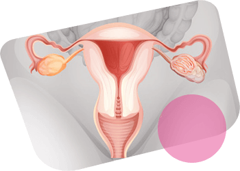
In DUB of reproductive age, ovarian persistence of follicles occurs with excessive secretion of estrogen (hyperestrogenemia).
The result of this is the absence of ovulation, corpus luteum, decreased secretion of progesterone and an increased risk of developing hyperplastic processes in the endometrium.
The diagnosis of DUB of reproductive age is made only after exclusion of diseases and pathological conditions in which uterine bleeding can also be observed: impaired uterine or ectopic pregnancy, residues of placental tissue, uterine fibroids with submucous or centripetal node growth, endometrial polyps, adenomyosis, polycystic ovaries, endometrial damage intrauterine contraceptives. Thus, the diagnosis of DUB is a diagnosis of exclusion. The main causes of bleeding are an imbalance of prostaglandins (PG's): the ratio of low PGF2a and high PGE2, increased fibrinolytic activity of the blood, impaired myometrial vascular structure, local defect in hormonal hemostasis in the endometrium.
One of the most common complications in women of any age with DUB is iron deficiency anemia. Given the negative consequences of anemia for a woman’s body, this problem should be recognized as one of the urgent in gynecology.
DUB - usually hormone-dependent bleeding
It is customary to distinguish ovulatory and anovulatory DUB. The key to differential diagnosis is determining hormonal status!
Ovulatory cycles, as a rule, occur with a regular menstrual cycle with a characteristic premenstrual syndrome (breast engorgement, dysmenorrhea), two-phase basal temperature.
Ovulatory DUBs make up about 20% of all DUB in women of reproductive age.
Ovulatory DUB is divided into intermenstrual and due to the persistence of the corpus luteum.
Intermenstrual DUB
Intermenstrual DUB is observed in the middle of the menstrual cycle; on days corresponding to ovulation, it lasts 2-3 days and is rarely intense. In their pathogenesis, the main role is played by a drop in the level of estrogen in the blood after the ovulatory peak of hormones. The differential diagnosis is carried out with polyps of the cervix or uterus, endometriosis of the cervix, erosion or cancer of the cervix.
DUB due to persistence of the corpus luteum
The persistence of the corpus luteum of the ovary is a consequence of a violation of the gonadotropic stimulation of progesterone synthesis. Its causes are not fully understood. Increased levels of progesterone in the blood and its prolonged secretion interfere with normal endometrial rejection during menstruation. Prolonged bleeding is facilitated by difficult rejection of the endometrium, a slowdown in reparative processes in it, as well as a decrease in the tone of the myometrium under the influence of an increased progesterone level in the blood.
Treatment of ovulatory uterine bleeding
Treatment of ovulatory uterine bleeding is usually conservative.
The effectiveness of the appointment of non-steroidal anti-inflammatory drugs (inhibitors of prostaglandin synthesis) has been proved: nimesulide, diclofenac, naproxen and tranexamic acid - antifibrinolytic (no more than 5 days). In order to prevent and reduce the recurrence rate, it is recommended to take low-dose combined oral contraceptives for 3-6 months. For women who are not planning a pregnancy in the coming years, the most effective levonorgestrel-releasing intrauterine system.
Anovulatory DUB
Anovulatory DUB occurs acyclic at intervals of 1.5-6 months, usually lasting more than 10 days. They are more common in women <20 and> 40 years old and are caused by hormonal imbalances in the hypothalamus-pituitary-ovary system. With this type of DUB, irregular anovulatory menstrual cycles, “breakthrough” bleeding, heavy menstruation, monophasic basal temperature are observed.
Causes of anovulatory bleeding: systemic hormonal imbalance, menstrual irregularities (oligo-, opsomenorrhea), lack of progesterone (in the luteal phase), which causes DUB, endometrial hyperplasia, infertility.
Anovulatory bleeding can also be caused by secondary hypothalamic diseases, such as stress, malnutrition, professional sports, weight loss, chronic systemic diseases, diseases of the endocrine system (thyroid gland, diabetes mellitus, adrenal hyperplasia, Cushing's syndrome, hyperprolactinemia), PCJ, obesity, hirsutism, insulin resistance.
It should also be remembered that DUB can occur while taking a number of medications: anticoagulants, antidepressants, combined oral contraceptives, tamoxifen, psychotropic, corticosteroids and chemotherapy. It is necessary to exclude liver diseases, coagulopathy and Willibrant disease.
Anovulatory bleeding treatment
The main objective of the treatment of anovulatory bleeding is the regulation of the menstrual cycle and the prevention of endometrial cancer.
With anovulatory bleeding, the first line of treatment is drug therapy. A 3-month course of combined oral contraceptives and progestogens in the 2nd phase of the cycle are recommended (10 mg dydrogesterone 2 times a day from the 11th to the 25th days of the cycle). Such a regimen reduces menstrual blood loss by 25–35% and does not have serious side effects. It is also possible to stimulate ovulation with clomiphene citrate at 50 mg / day from the 5th to the 9th days of the cycle. In cases where the above extragenital diseases are the cause of DUB, their pathogenetic correction is necessary.
The diagnosis of DUB of reproductive age is made only after exclusion of diseases and pathological conditions in which uterine bleeding can also be observed: impaired uterine or ectopic pregnancy, residues of placental tissue, uterine fibroids with submucous or centripetal node growth, endometrial polyps, adenomyosis, polycystic ovaries, endometrial damage intrauterine contraceptives.
If hormonal treatment is ineffective, separate diagnostic curettage under the control of hysteroscopy is indicated. Ablation (hysteroresectoscopy, etc.) of the endometrium is preferable for women of late reproductive age who have realized a generative function.
In the absence of the effect of conservative treatment methods, surgical treatment is indicated - hysterectomy (in exceptional cases).
Premenopausal DUB
DUB in the premenopausal period in women 45–55 years of age is the most common gynecological pathology. These bleeding occur due to age-related changes in the functional state of the hypothalamic structures that regulate the function of the ovaries. First of all, the cyclical release of luliberin and, accordingly, FSH and LH is disrupted, as a result of which the ovarian function is impaired: anovulation and a lack of progesterone. Deficiency of progesterone against the background of relative hyperestrogenemia leads to the same changes in the endometrium as in DUB of the reproductive period: hyperplastic processes, such as atypical hyperplasia and endometrial adenomatosis, occur more frequently in premenopause than in reproductive age. At the same time, age-related immunosuppression further increases the risk of developing endometrial malignancies. Premenopausal DUB is often combined with adenomyosis (20%), uterine myoma (25%), and endometrial polyps (30%). Hormone-active ovarian tumors can be a relatively rare cause of DUB and recurrent endometrial processes.
One of the most common complications in women of any age with DUB is iron deficiency anemia. Given the negative consequences of anemia for a woman’s body, this problem should be recognized as one of the urgent in gynecology.
The condition of patients, as well as with DUB of other age periods, is determined by the degree of hypovolemia and anemia, but, given the high frequency of concomitant diseases and metabolic endocrine disorders (arterial hypertension, obesity, diabetes mellitus, etc.), DUB in women 45–55 years are more difficult than in other age periods.
The main therapeutic measure is a separate diagnostic curettage of the mucous membrane of the cervix and uterine body under the control of hysteroscopy, followed by histological examination of the material obtained by curettage. The use of conservative hemostasis with hormonal drugs before curettage is a gross medical error. In the future, the tactics of treatment of DUB is determined by the presence of concomitant gynecological pathology, diseases of other organs and systems, the age of the patient. An absolute indication for removal of the uterus is a combination of DUB with recurrent adenomatous or atypical endometrial hyperplasia, a nodular form of uterine adenomyosis, uterine myoma with a submucous node location. A relative indication for surgical treatment is the combination of DUB with recurrent glandular-cystic endometrial hyperplasia in women with obesity, clinically expressed diabetes mellitus, and arterial hypertension.
Biofer is a stable complex of ferric iron and maltose, which has high structural homogeneity and is able to easily deliver its constituent iron to endogenous iron-binding proteins.
Iron deficiency anemia is a disease in which the iron content in the blood serum and bone marrow decreases. Diagnosis of iron deficiency anemia is not particularly difficult. It is indisputable that in order to combat iron deficiency it is necessary to eliminate its root cause using special iron preparations. Some modern preparations of ferrous iron in the form of sulfate, fumarate and succinate can cause side effects (irritation of the stomach, nausea, etc.). The use of ferric iron preparations instead of the ferrous iron preparations mentioned above is very promising. These drugs include Biofer, a stable complex of ferric iron and maltose, which has high structural homogeneity and is able to easily deliver its iron to endogenous iron-binding proteins. A comparative study showed that in patients who do not tolerate ferrous succinate, the iron (III) complex polymaltosate hydroxide + folic acid successfully eliminates iron deficiency and rarely causes side effects. In addition, it was previously established that with oral administration of iron (III), polymaltosate hydroxide simultaneously with food does not change iron absorption, and the use of a complex labeled with 59Ре (III) revealed a significant increase in absorption.
Treatment with Biofer should be long. The standard dosage is 1 tablet 2-3 times a day. The effectiveness of therapy is assessed on the 9-12th day of treatment by counting the number of reticulocytes and comparing them with the initial level. Hemoglobin levels usually increase by the end of the 3rd week of therapy, and the red blood cell count after 5-8 weeks of treatment. Normalization of the hemoglobin content in iron deficiency anemia does not serve as a basis for discontinuing treatment. To replenish iron reserves in the body, long-term maintenance therapy with small doses is necessary based on the dosage of Biofer, 1 tablet 1 time per day. If blood loss remains intense and the hemoglobin content drops after a six-month or one-year break in treatment, monthly courses of treatment with iron preparations are necessary 1-2 times a year in therapeutic doses.
Given the high efficiency and good tolerability of the drug, for the treatment of posthemorrhagic anemia in patients with DUB, it is possible to recommend a polymaltose iron complex in an amount equivalent to 100 mg of elemental iron and folic acid 350 μg (chemical composition of Biofer).
Treatment of DUB depends on the age of patients, which are divided into 3 groups: younger than 20 years old, between 20 and 40 years and older than 40 years.
Treatment goals:
- Treatment of the root cause.
- Restoration and regulation of the menstrual cycle, ovulation and the full luteal phase of the cycle.
- Prevention of bleeding and reduction of future risks.
- Normalization of hormonal parameters.
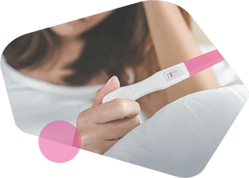
- The onset of pregnancy.
- Improving the quality of life.
- In the absence of pregnancy - determination and monitoring of the endometrium and early detection of neoplastic processes of endo- and myometrium.
- Examination and treatment of patients should be individual.