

Anatomy and physiology of the female reproductive system
The structure of the female genital organs
Among the female genital organs, external and internal are distinguished. To the outside belong the pubis, labia majora, labia minora, clitoris, vestibule of the vagina. The hymen is the boundary between the external and internal genital organs. The external genitalia of women vary greatly in appearance. The differences relate to the size, shape and pigmentation of the labia, color, texture, amount and distribution of pubic hair, the appearance of the clitoris, vestibule and hymen. The genitals of different people vary in their structure in the same way as the structure of their faces.
Female internal genital organs
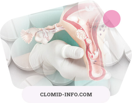
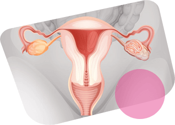
Internal genitalia include: the vagina, uterus, uterine appendages (fallopian tubes and ovaries). Ligaments that suspend the uterus and appendages can also be considered internal genital organs. Internal genital organs are located inside the pelvic ring.
Vagina
The vagina (vagina) is a whole-tissue channel with a length of 7-8 to 9-10 cm. It is attached to the place of transition of the cervix into its body. Here the cervix protrudes into the lumen of the vagina (the vaginal part of the cervix). In the place of attachment of the vagina to the cervix, the arch is obtained: front, back, left and right. The least deep is the anterior arch, the deepest is the posterior. The vagina is an internal organ formed by muscle tissue and located diagonally at an angle of 45 ° to the lower back.
In the absence of sexual stimulation, the vaginal walls collapse. In a nulliparous woman, the length of the back wall of the vagina is on average 8 cm, and the front - 6 cm.
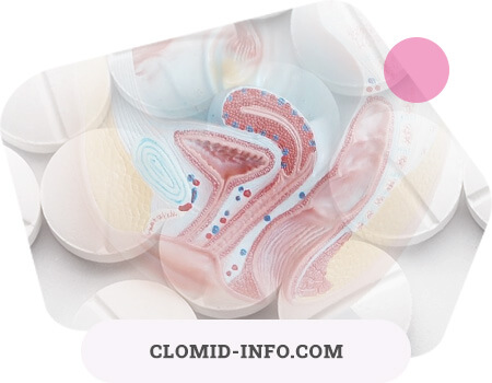
The mucous membrane consists of a stratified squamous epithelium, there are no glands in it. Epithelial cells contain glycogen, lactic acid is obtained from it, which determines the optimal conditions for the existence of non-pathogenic bacilli, the so-called vaginal rods (Doderlein rods). The acidic environment of the vaginal contents and the presence of sticks prevent the development of pathogenic microbes.
During sexual arousal, blood plasma (so-called “sweating”) is released into the lumen of this penis through the walls of the venous vessels of the vagina, which, when mixed with the secret of the bertoline glands, form a “lubricant” that facilitates gliding of the penis.
The average length of the vagina in an unexcited state is 8-12 cm, but thanks to the muscles and folds, when excited, the vagina can be very stretched both in length and in width, tightly covering the male genital organ of almost any size. Therefore, the size of the penis practically does not affect the female orgasm.
According to some anatomists, at a depth of several centimeters in the vagina there is the so-called "point - G", the vaginal zone, comparable in sensitivity to the clitoris. However, it should be remembered that most scientists consider the existence of a similar part of the genital organs in a woman unproven. Therefore, it is hardly worth focusing on the search for this point during intercourse.
The vagina, like an inflatable balloon, can change its shape and size. It is able to expand, creating conditions for the passage of the baby’s head during childbirth, or shrink enough to cover the finger introduced into it from all sides.
Despite its ability to contract, a woman’s vagina cannot cover the penis during intercourse so tightly that physical separation becomes impossible. Bonding, which sometimes occurs in dogs, is mainly due to the expansion of the bulbar part of the penis.
Many people are interested in the relationship between vaginal size and sexual satisfaction. Since the width of the vagina adapts equally well to a large or small penis, a mismatch in the size of the genital organs of men and women is rarely the cause of complications in sexual relationships. After birth, the vagina usually expands somewhat and its elasticity decreases to some extent. According to some authors, in such cases, exercises can help to strengthen the muscles supporting the vagina, which will increase sexual reactivity.
“Cagel (Kegel) exercises” consist in contracting the pelvic muscles that support the vagina, namely the bulbous cavernous (bulbocavernosus) and pubicoccygeal (pubo coccygeus). These same muscles contract when a woman stops urinating or compresses the vagina, preventing the introduction of a tampon, finger or penis. During exercise, the muscles contract strongly for one to two seconds, and then relax; to achieve maximum results, such reductions should be repeated several times a day, making 10 reductions each time. In addition to strengthening muscles, these exercises allow a woman to know herself. However, it is currently not entirely clear whether sexual reactivity is increasing.
The inner lining of the vagina is similar to the oral mucosa. The vaginal mucosa provides moisture. There are no secretory glands in the vagina, but it is rich in blood vessels. The ends of the sensory nerve fibers are at the entrance to the vagina, and in the remaining parts of them there are relatively few. As a result, the deeper part of the vagina (approximately two-thirds) is relatively less sensitive to touch or pain.
In recent years, disputes over the existence on the front wall of the vagina (halfway between the pubic bone and cervix) of a certain area, especially sensitive to erotic stimulation, have not subsided. This area, called zone G (after the name of the German physician Grefenberg, who described it in 1950), in an unexcited state has the size of an ordinary bean, but with stimulation it greatly increases due to tissue swelling.
Ladas, Whipple and Perry (1982) argue that by examining more than 400 women, they found zone G in each of them; in their opinion, this structure had previously gone unnoticed, because "in the absence of excitation, it is very small and difficult to detect." These data contradict the results of studies in which Whipple herself later participated: zone G was detected only in 4 out of 11 women; its existence is not confirmed by the data of our studies conducted at the Masters and Johnson Institute: of the 100 carefully examined women, only 10% had an area of hypersensitivity on the front wall of the vagina or a lump of compacted tissue corresponding to the descriptions of zone G. Similar studies also did not reveal the presence of zone G, although many women have noted increased erotic sensitivity on the front wall of the vagina. In later works, the conclusion was drawn that "the presence of zone G ... even among a minority of women, not to mention their majority, cannot yet be considered proven." Thus, additional studies are needed to establish whether zone G really exists as a separate anatomical structure, or, as Helen Kaplan writes, “the idea that many women have special erogenous zones in the vagina that enhance pleasure and orgasm is not new and should not be controversial. "
Perhaps a higher sensitivity of the anterior vaginal wall is "an integral part of the clitoris orgasmic reflex."
The lower part of the uterus - the cervix (cervix) protrudes into the vagina. From the side of the vagina, the neck of a nulliparous woman looks like a smooth pink button with a rounded surface and a small hole in the center. Sperm penetrate the uterus through the pharynx of the cervix (cervical os); through it, menstrual blood is released from the uterus. The cervical canal (a thin tube connecting the pharynx of the cervix to the uterine cavity) contains numerous glands that produce mucus. The consistency of this mucus depends on the hormonal background and therefore changes at different stages of the menstrual cycle:
- immediately before ovulation or in the process of the latter (when the egg leaves the ovary), the mucus becomes liquid and watery;
- at other times, it is thick and forms a cork blocking the entrance to the cervix.
There are no superficial nerve endings in the cervix, and therefore touching it almost does not cause sexual sensations; surgical removal of the neck does not reduce the sexual activity of a woman.
Uterus
The uterus (uterus) is a hollow muscle organ that has the shape of an inverted upside down and somewhat flattened pear.
Its length is approximately 7.5 cm and a width of 5 cm. Anatomically, the uterus is divided into several parts. The endometrium lining the uterus from the inside and its muscle component, the myometrium, perform different functions. During the menstrual cycle, the endometrium undergoes changes, and at the beginning of pregnancy, a fertilized egg is implanted in it.
The muscular wall is actively involved in childbirth and delivery. Both functions of the uterus are regulated by hormones - chemicals that also cause an increase in the uterus during pregnancy. The uterus is fixed in the pelvic cavity using six ligaments, but not very rigidly.
The angle between the uterus and the vagina varies in different women. Typically, the uterus is located more or less perpendicular to the axis of the vaginal canal, but in about 25% of women it is bent backward, and in about 10% - forward. Sometimes this anatomy of the internal genital organs can cause pain during intercourse during deep frictions, since the head of the penis can hit the outer walls of the uterus. In this case, you need to choose the position of sexual intercourse, in which the male genital organ does not enter the vagina to the full depth.
Since the nerve endings on the genitals of a man are maximally concentrated on the head of the penis, and in a woman - in the lower part of the vagina, such postures do not affect the intensity of sensations in both partners.
In cases where the uterus is rigidly fixed by adhesions that occur after operations or as a result of the inflammatory process, a woman may feel pain during intercourse; this situation requires surgical intervention.
Isthmus
The isthmus is a canal about 1 cm long, located between the uterine cavity and the cervical canal. On the site of the isthmus is located the internal pharynx of the cervix. During pregnancy and childbirth, the lower body of the uterus and the isthmus make up the lower segment of the uterus.
The cervix partially protrudes into the lumen of the vagina (vaginal part), partially is located above the vagina (supravaginal part). In women who have not given birth, the cervix has a conical shape. In women who gave birth, the cervix is wider and has a cylindrical shape. The cervical canal (cervical canal) is also cylindrical in shape. The external opening of the cervical canal is called the external pharynx. For those who did not give birth, it is roundish, "point", and for those who have given birth, it is slit-like due to lateral ruptures of the neck during childbirth.
Sperm enter the uterus through the cervical canal, and during menstruation, discharge comes out. During sexual arousal, the uterus rises, lengthening the vagina.
The fallopian tubes
The fallopian tubes (fallopian tubes) are narrow tubes with a pronounced muscle layer that are constantly contracting. Their mucous membrane consists of cells with cilia, which create a flow of fluid in the direction from the pelvic cavity to the uterine cavity. Thus, the egg is transported from the ovary to the uterus. On the way - in the tube - the egg is fertilized - it merges with the sperm. The egg becomes heavier and more slowly gets to the uterine cavity. Violation of the ciliary apparatus due to inflammation of the tube, narrowing of the tube, violation of the agreed muscle contraction leads to the fact that the egg settles in the tube and an ectopic tube pregnancy develops.
The length of the fallopian tubes is about 10 cm. The tube consists of four parts: intramural (passes through the wall of the uterus), the isthmus (the narrowest section of the tube next to the uterus), the ampulla (the longest tortuous part of the tube), the abdominal (end) that opens with the funnel in abdominal cavity.
Unlike men, in whom the abdominal cavity is isolated from the external environment, in women, the abdominal cavity is connected to the external environment. Thus, women are more likely to get infection through the genitals into the abdominal cavity. The fallopian tubes are also called oviducts, because the eggs move along the canal from the abdominal cavity to the uterine cavity.
Ovaries
The ovaries (ovaries), or female gonads, are paired organs located on both sides of the uterus. Larger ovaries can be compared with inshell almonds (approximately 3 x 2 x 1.5 cm); they are held in place by connective tissue, which attaches to the wide ligament of the uterus.
Even before the birth of a girl, the development of future eggs begins in her forming ovaries. At about 5-6 months of pregnancy, the ovaries of the fetus contain 6-7 million future eggs, most of which are atresia before the birth of the girl. The ovary of the newborn contains approximately 400,000 immature eggs; no new eggs are formed in the future. In childhood, atresia continues, and the number of eggs decreases even more. Immature oocytes are surrounded by a thin layer of cells that form the follicle.
The ovaries are the female reproductive glands (paired organ). They are located in a separate deepening of the peritoneum and are attached to the posterior wall of the peritoneum with a wide ligament. The size of the ovary is 3 x 2 x 1 cm, and it weighs about 7 g. The main layer of the ovary is the cortical substance, which covers the inner layer - the medulla. In the cortical layer follicles are placed, in which there are eggs. In the cerebral layer, which consists of a softer connective tissue, numerous blood and lymph vessels, nerves pass. The ovaries perform two functions: they produce hormones (the most important of them are estradiol and progesterone) and they produce eggs.
The fallopian tubes, ovaries, and uterine ligaments are called uterine appendages.
The normal, typical location of the internal genital organs is facilitated by the own tone of the genital organs, the coordinated activity of the diaphragm, abdominal press and pelvic floor, as well as the ligamentous apparatus of the uterus.
Pelvic cavity peritoneum
In women in the pelvic cavity, the parietal sheet of the peritoneum, having descended from the abdominal cavity along its posterior wall, passes through linea terminalis, covering the meso-peritoneal anterior surface of the middle third of the rectum. Then the peritoneum passes to the posterior vaginal fornix and, following up, covers the posterior surface of the uterus, reaching its bottom. Here, the peritoneum again lowers and covers the front surface of the uterus, reaching its neck. Moving further to the posterior surface of the bladder, it follows upward, reaches its apex, and then passes into the parietal peritoneum lining the inner surface of the anterior abdominal wall. Thus, with respect to the uterus, the peritoneum forms two depressions located in the frontal plane: one between the rectum and the uterus - the rectal-uterine depression, excavatio rectouterina, and the second between the uterus and the bladder - the vesicoureteral cavity, excavatio vesicouterina. The first deepening is much deeper and is limited along the edges of the rectal-uterine folds, plicae rectouterinae, in the thickness of which are underdeveloped muscles of the same name containing smooth muscle fibers. The second deepening, excavatio vesicouterina, is less than the first; its depth depends on the degree of filling of the bladder. Both recesses, except for the uterus, are separated from one another by its wide ligaments, ligg, lata uteri, which are the duplication of the peritoneum.
Blood supply to the external genital organs occurs due to the shameless artery and, in part, the branches of the femoral artery. The internal genital organs are supplied with blood through the hypogastric artery, branches of the uterine and vaginal arteries, as well as through the ovarian artery. Outflow of venous blood occurs along the veins of the same name.
The lymphatic system is a network of sinuous lymphatic vessels and lymph nodes located along the blood vessels in the direction of movement of the venous blood.
The nervous system consists of the sympathetic and parasympathetic parts, as well as spinal nerves. In the innervation of the genital organs, the solar, hypogastric and utero-vaginal (or pelvic, sacral) plexuses take part. Sensitive nerve endings from the genitals are connected with the subcortical nerve centers and with the cerebral cortex and form a single complex system for regulating physiological processes in the genital apparatus, including the development of these organs, menstrual and reproductive functions, and the period of extinction (menopause).
Articles


Female external genitalia
Pubis
The pubis (mons veneris) is an eminence, consisting of adipose tissue, located in front and slightly above the pubic joint, covered with skin and hair, the upper boundary of the growth of which is horizontal (unlike men whose hair growth extends upward along the midline).
There are many nerve endings in this area, and therefore touching and / or pressing can cause sexual arousal. Many women believe that pubic stimulation causes the same pleasant sensations as direct contact with the clitoris.
Labia minora
The labia minora is located deeper, behind the labia minora. In front, they seem to exit the clitoris, forming two legs that go back. The labia minora is covered by a thin layer of skin that resembles a mucous membrane of a pale pink color. If the small lips protrude beyond the borders of the large, then the skin that covers them is dark brown.
The labia minora are like curved petals. Their core is formed by a spongy tissue rich in small blood vessels and does not contain fat cells. The skin covering the labia minora is devoid of hair but contains many nerve endings. Small lips converge over the clitoris, forming a skin fold called the clitoris foreskin. This area of the labia minora is sometimes called the female foreskin.
For many women, the labia minora is one of the main erogenous zones. The tissue forming the labia minora does not contain fat, but is penetrated by venous vessels, resembling the cavernous bodies in the genitals of a man. When excited, the labia minora fill up with blood and swell somewhat. When the skin covering the labia is infected, intercourse can become painful, itching or burning can also occur.
On the inner surface of the labia minora there are ducts of the so-called bertolin glands (two paired glands that produce mucus during sexual arousal, which facilitates penetration of the penis into the vagina, the glands themselves are located in the thickness of the labia majora). It was once believed that these glands play a major role in the production of vaginal lubrication, but it has now been established that the few drops of secretion that they usually secrete during sexual arousal only moisturize the labia slightly.
Large labia
Large labia majora (labia majora) are pronounced longitudinal folds of skin located on the sides of the genital fissure, under which there is a subcutaneous base with fibrous fibers, where blood vessels and nerves pass and the bartholin glands are placed. The large labia in front converge into the anterior commissure, which is located above the clitoris and covers it. Back, the labia majora narrow and converge to one another and pass into the posterior commissure. The skin of the outer surface of the labia majora is covered with hair; sweat and sebaceous glands are located in it. On the inside, the labia majora is covered with thin pink skin that looks like a mucous membrane. The genital gap is the space between the labia majora.
The skin of the labia majora has many nerve endings. Although, only a small percentage of women stimulation of the labia majora causes excitement. In the absence of sexual stimulation, the labia majora are usually closed in the midline, which creates mechanical protection for the opening of the urethra and the entrance to the vagina.
Crotch
Crotch is the space between the posterior commissure of the labia majora and the external opening of the anus. Outside, the crotch is covered with skin, on which the line from the posterior commissure to the anus is visible - the crotch seam. In the thickness of the perineum are three layers of muscles that make up the pelvic floor. The distance from the posterior commissure to the anus is called the height of the perineum; it is 3-4 cm. With a high or low elongated (rigid) perineum, during childbirth, in order to avoid tearing of the perineum, it is cut (episiotomy).
This area is often sensitive to touch, pressure, temperature and can be a source of sexual arousal.
Clitoris
The clitoris is a small cone-shaped formation that consists of cavernous bodies, similar to the structure of the male penis. In the cavernous bodies there are connected voids filled with circulating blood, which comes here from the blood vessels. With sexual arousal, the clitoris is intensely filled with blood, it increases and hardens (erection), since the clitoris has many vessels and nerves. The cavernous bodies are not capable of contractions and cannot be completely thrombosed, therefore traumatic damage to the clitoris is dangerous.
The clitoris is the most mysterious part of the female reproductive system, the most unknown, the most necessary in the sex life.
The clitoris, one of the most sensitive areas of the female genital organs, is located where the tips of the labia minora converge.
The clitoris head resembles a small shiny button. To see it, one must carefully push the foreskin (skin) covering the clitoris.
The body of the clitoris (corpus clitoris) consists of a spongy tissue that forms two long legs (crura) in the form of an inverted letter V.
The legs are directed to the pelvic bones. The clitoris is rich in nerve endings, which makes it very sensitive to touch, pressure and temperature. This is a unique organ, the only function known to us of which is to concentrate and accumulate a woman's sexual sensations.
The clitoris is often regarded as a miniature penis, however this is a sexually colored and misconception. The clitoris is neither involved in reproduction nor in urination; it does not lengthen, unlike the penis, during stimulation, although it also overflows with blood.
In the process of embryonic development, the clitoris and penis are formed from the same embryo.
The size and appearance of the clitoris vary greatly, but there is no evidence that large clitoris can create stronger sexual arousal
It is believed that circumcision of the clitoris - the surgical removal of the foreskin - enhances a woman's sexual reactivity, since it becomes possible to stimulate the clitoris head more directly. However, this practice can only help.
in rare cases, since it has two major drawbacks:
- the clitoris head is often too sensitive to direct touch, which sometimes even causes pain or irritation (in this sense, the foreskin has a protective function)
- during intercourse, the introduction of the penis into the vagina indirectly stimulates the clitoris, moving the labia minora, as a result of which the foreskin rubs against the head of the clitoris.
Some tribes in Africa and South America practice surgical clitoral removal (clitorectomy) as a ritual ceremony after puberty. According to one doctor in Egypt, some young girls are still undergoing this painful procedure.
Although this operation is called "clitoris circumcision", in fact it is not at all that. Clitorectomy does not violate sexual arousal or orgasm, but does not contribute to their strengthening.
It is for this reason that most women during masturbation only stroke the area around the head of the clitoris, avoiding its direct stimulation. Apparently, supporters of clitoral circumcision (oddly enough, these are usually men) did not pay due attention to this circumstance.
The labia minora in its upper junction form the foreskin and frenum of the clitoris.
In a calm state, the clitoris head is almost invisible under the bridle. However, with excitation, an erection of the clitoris occurs, and this genital organ can significantly increase in size, speaking over the bridle. However, the degree of clitoris enlargement upon excitation varies greatly among different women. Also, it should be noted that the erection of the clitoris is much slower than the penis in men. In order for the clitoris to increase in size, it takes time from 20 seconds to several minutes.
Clitoral enlargement occurs in proportion to the degree of arousal, however, immediately before orgasm, the clitoris again decreases in size (this is one of the signs of orgasm in a woman), then swells again.
Due to the high sensitivity, this part of the genitals of a woman should be stimulated carefully (in some women, direct stimulation of the clitoris causes negative sensations, due to the very high sensitivity of this genital organ). Basically, to excite a woman and achieve her orgasm, it is enough to just gently stroke this genital organ. In addition, it should be remembered that the caress of the clitoris can begin only after moisturizing the genitals.
Do not forget that after an orgasm, touching the clitoris in most women causes discomfort.
Vaginal vestibule
The border of the vestibule is the hymen or its remnants that separate the external genitalia from the internal. In front, the vestibule is limited by the clitoris, behind - by the rear commissure, on the sides - by the labia minora. Under the clitoris is the external opening of the urethra. The excretory ducts of the large glands of the vaginal vestibule are located on the sides and below the urethral opening.
Onion vestibule
The bulb of the vestibule (bulbus vestibuli), corresponds to the bulb of the penis, but has a number of differences. The bulb is an unpaired formation consisting of two - right and left - parts that are connected by a small intermediate part located between the clitoris and the external opening of the urethra. Each lobe is a dense venous plexus, in which elongated lateral parts are laid at the base of the labia majora; they represent a flattened, spindle-shaped form of formation, which, thickening posteriorly, cover the large glands of the vestibule with their rear end. Outside and below, each of the halves of the vestibule is covered by a bulbous-spongy muscle, w. bulbospongiosus. The vestibule bulb has a protein membrane encircling the venous plexus, which is penetrated by smooth muscle fibers and connective tissue bundles.
Urethra
The urethra has a length of 3-4 cm, its lumen is stretched to 1 cm or more. The external opening of the urethra is round, half a month, or star-shaped, located 2-3 cm below the clitoris. The urethra is connected to the front wall of the vagina throughout. On both sides of the urethra, there are external openings of the paraurethral passages (or skin sinuses) that are 1-2 cm long. A secret is produced in these formations that moisturizes the area of the external opening of the urethra.
Large glands of the vestibule
The large glands of the vestibule are an oblong-round formation the size of a bean, of a densely elastic consistency, located on the border of the posterior and middle third of the labia majora. In the alveoli of the glands, a secret is produced. Excretory ducts of the large glands of the vestibule (Bartholin glands) open from the inside of the labia minora at the level of the location of the Bartholin glands. The secret of the large glands of the vestibule has a whitish color, an alkaline reaction, a specific smell. It is secreted during sexual intercourse and helps to moisturize the vagina.
Hymen
Hymen (hymen) is a whole-tissue membrane covered on both sides by a stratified squamous epithelium. It most often has one, sometimes several holes. With the onset of sexual activity, the hymen ruptures. The hymen usually has openings through which blood is released during menstruation. The left side does not completely cover the entrance to the vagina and varies in shape, size and thickness.
An annular pleura surrounds the opening of the vagina; cloisonne pleura consists of one or more strips of tissue crossing the opening of the vagina; the ethmoid pleura completely tightens the vaginal opening, but there are many small holes in it; parous introitus (opening of the vagina of the woman giving birth) - only the remains of the hymen are visible.
In earlier times, a girl who entered into marriage should have had a hymen undisturbed, which served as evidence of her innocence. The bride, whose hymen was torn, could be returned to parents, subjected to public ridicule or corporal punishment, and in some countries even sentenced to death. Nowadays, brides who want to hide past sexual relationships from their future husbands are turning to a doctor to restore hymen with the help of plastic surgery.
Contrary to the opinion of most women, the doctor, conducting a gynecological examination, can not always say whether the patient is a virgin. The integrity or disturbance of the hymen cannot be considered a solid sign of a woman's sexual behavior in the past. The lobe could be torn or stretched in early childhood as a result of various exercises or the insertion of fingers or any objects into the vagina. In some women, the pleura from birth covers the entrance to the vagina only partially or is completely absent. On the other hand, sexual intercourse does not always lead to a rupture of the hymen; sometimes it just stretches. In most cases, the first sexual intercourse is not painful and is not accompanied by severe bleeding. The excitement associated with the event is usually quite large, and the pressure exerted on the hymen is not enough to violate its integrity.